Difference between revisions of "Publications/xu.17.icip.inc"
From LRDE
Yongchao Xu (talk | contribs) |
Yongchao Xu (talk | contribs) |
||
| Line 106: | Line 106: | ||
File:Xu.17.icip-axialTest430Input.png |
File:Xu.17.icip-axialTest430Input.png |
||
File:Xu.17.icip-axialTest440Input.png |
File:Xu.17.icip-axialTest440Input.png |
||
| + | </gallery> |
||
| − | |||
| + | <gallery> |
||
| − | |||
File:Xu.17.icip-axialTest408Seg.png |
File:Xu.17.icip-axialTest408Seg.png |
||
File:Xu.17.icip-axialTest418Seg.png |
File:Xu.17.icip-axialTest418Seg.png |
||
| Line 124: | Line 124: | ||
File:Xu.17.icip-coronal30Test529Input.png |
File:Xu.17.icip-coronal30Test529Input.png |
||
File:Xu.17.icip-coronal30Test540Input.png |
File:Xu.17.icip-coronal30Test540Input.png |
||
| + | </gallery> |
||
| − | |||
| + | <gallery> |
||
| − | |||
File:Xu.17.icip-coronal30Test509Seg.png |
File:Xu.17.icip-coronal30Test509Seg.png |
||
File:Xu.17.icip-coronal30Test517Seg.png |
File:Xu.17.icip-coronal30Test517Seg.png |
||
| Line 142: | Line 142: | ||
File:Xu.17.icip-coronal40Test5070Input.png |
File:Xu.17.icip-coronal40Test5070Input.png |
||
File:Xu.17.icip-coronal40Test5094Input.png |
File:Xu.17.icip-coronal40Test5094Input.png |
||
| + | </gallery> |
||
| − | |||
| + | <gallery> |
||
| − | |||
File:Xu.17.icip-coronal40Test5021Seg.png |
File:Xu.17.icip-coronal40Test5021Seg.png |
||
File:Xu.17.icip-coronal40Test5054Seg.png |
File:Xu.17.icip-coronal40Test5054Seg.png |
||
Revision as of 14:38, 6 February 2017
Method and datasets
Method
Architecture of the proposed network. We fine tune it and combine linearly fine to coarse feature maps of the pre-trained VGG network. The coarsest feature maps are discarded for the adult images.

Datasets
- Dataset of the MICCAI challenge of Neonatal Brain Segmentation 2012 (NeoBrainS12)
- Axial images acquired at 40 weeks: 2 training images + 5 test images
- Coronal images acquired at 30 weeks: 2 training images + 5 test images
- Coronal images acquired at 40 weeks: 5 test images
- Dataset of the MICCAI challenge of MR Brain Image Segmentation (MRBrainS13)
- Axial images acquired at 70 years: 5 training images + 15 test images
Materials
Illustrations
Experiments
Leave-One-Subject-Out (LOSO) cross-validation on N images + normal training/test experiments. Note that only one training image is used for LOSO 2.

LOSO experiments
Quantitative results of LOSO experiments in terms of Dice coefficient as compared to the state-of-the-art results. The last one is from P. Moeskops et al. on the 15 test images in MRBrainS13 dataset.

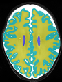
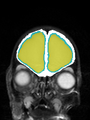
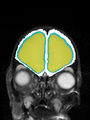
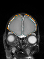
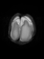
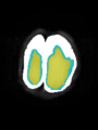
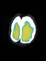
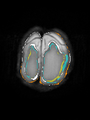
- Qualitative results on axial images at 40 weeks in NeoBrainS12 dataset
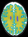
Input
Segmentation
Manual segmentation
Difference


- Qualitative results on coronal at 30 weeks in NeoBrainS12 dataset
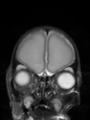
Input
Segmentation
Manual segmentation
Difference


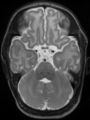
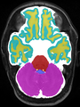
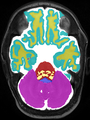
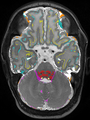
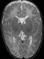
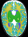
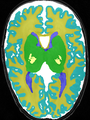
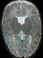
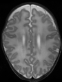
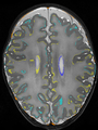
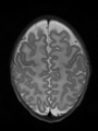
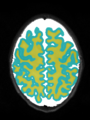
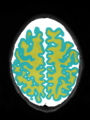
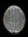
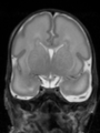
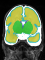
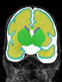
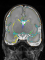
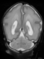
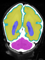
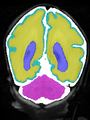
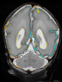
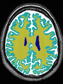
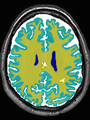
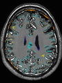
- Qualitative results on aging adult at 70 ages in MRBrainS13 dataset
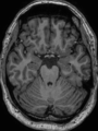
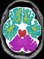
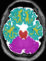
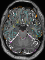
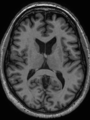
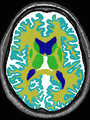
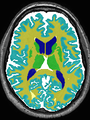
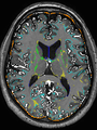
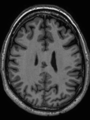
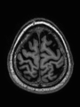
Input
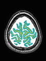
Segmentation
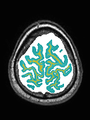
Manual segmentation
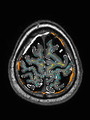
Difference


Neonatal brain MR image segmentation
- Results on axial images at 40 weeks in NeoBrainS12 dataset. More details can be found Here

Some qualitative results


- Results on coronal images at 30 weeks in NeoBrainS12 dataset. More details can be found Here

Some qualitative results


- Results on coronal images at 40 weeks in NeoBrainS12 dataset. More details can be found Here

Some qualitative results


Adult brain MR image segmentation
- Results on aging adult images at 70 years in MRBrainS13 dataset. Only top 10 methods among 38 submitted ones are shown. More results and details can be found Here

Some qualitative results


